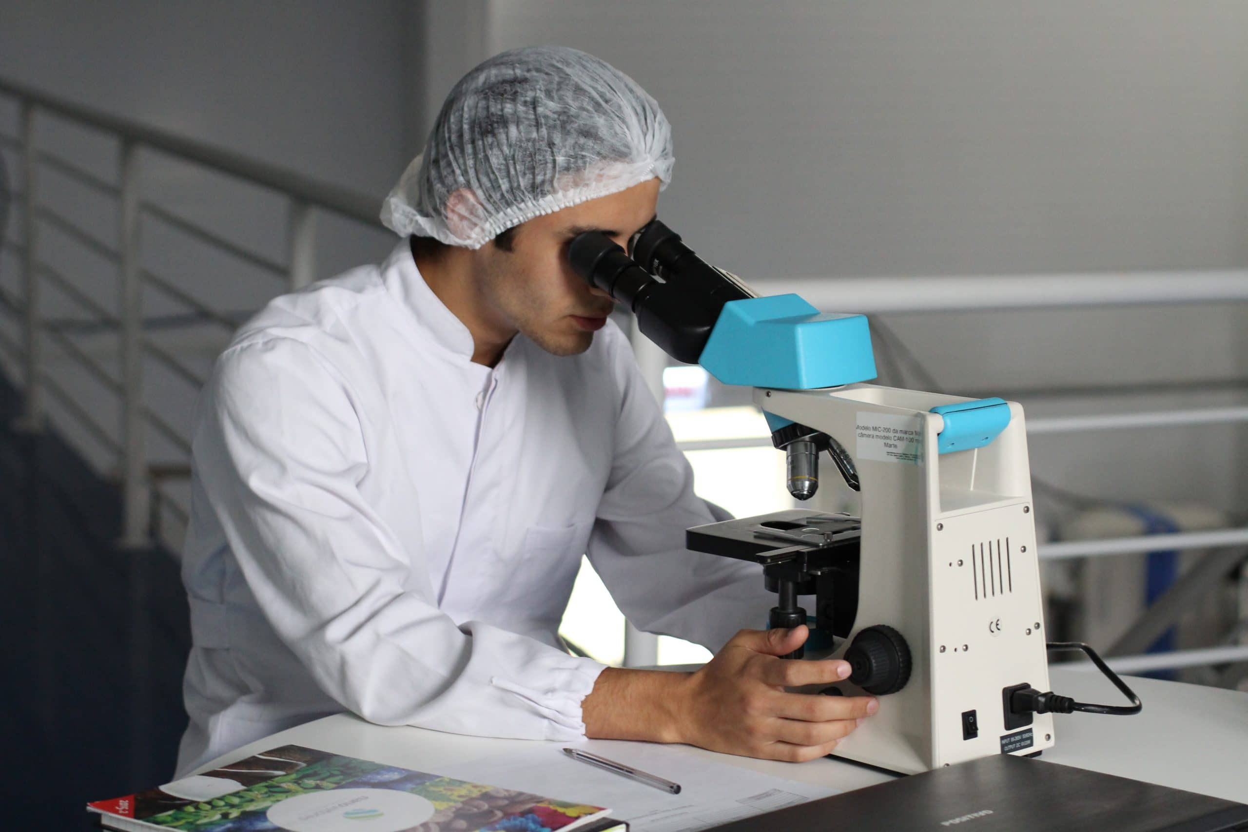
Within the field of material characterization, microscopy techniques are one of the most common tools in chemical laboratories. In this post we review which are the most common modalities, how they work and how they are applied in the industry today.
What are microscopy techniques?
Microscopy techniques are the set of research procedures that use a microscope to obtain images of certain structures, which, because they are too small, are invisible to the naked eye. Due to their great potential to identify material properties, they are used for a variety of purposes, ranging from molecular microbiology to the analysis of metals and other industrial compounds.
Main microscopic techniques for characterization of materials
There are two large groups of microscopic techniques for characterization of materials: optical microscopy and electron microscopy. In the first, the microscopes use photons of light to amplify the samples, while in the second, the images are generated by the interaction between electrons and different substances. By using particles with a shorter wavelength, electron microscopy has greater resolving power.
In addition, there is also a need for scanning probe microscopy, which produces images using a probe that travels the surface of the samples. Next, we review the most common microscopic techniques for characterizing materials:
Advanced Optical Microscopy (MOA)
It is the evolution of the traditional microscopic technique, in which objects are magnified using optical lenses and a beam of light. Today, advanced optical microscopy (MOA) uses equipment such as confocal microscopes, with nanometric resolution that provides detailed information on the shapes of materials and their texture.
Scanning electron microscopy (SEM)
The techniques of characterization of materials by means of scanning electron microscopy work with electromagnetic lenses, deflector coils, radiation detectors and a moving and concentrated electron beam that goes through the entire sample point by point. The resulting image is displayed on a screen and includes morphological, crystallographic, topographic, and compositional information.
Transmission electron microscopy (TEM)
Transmission electron microscopy differs from scanning in that it measures most of the samples at the same time (not point by point) and builds the image from the electrons that pass through the analyzed object, and not from those that are scatter or bounce off the surface. Additionally, TEM uses finer samples and has more powerful, atomic-level resolution.
Scanning Transmission Electron Microscopy (STEM)
This technique incorporates additional electromagnetic coils with which it launches a concentrated beam of electrons that sweeps the sample pixel by pixel, but unlike SEM it generates the image with the electrons that pass through the substances. The loss of energy enables the characterization of the chemical composition of the materials.
Tunnel effect microscopy (STM)
Tunneling microscopes operate with a fine-tipped probe to which a certain voltage is applied. As it approaches the surface of the sample, an exchange of electrons occurs between the closest points of the material and the edge of the probe. In this way, the relief of the surface is reconstructed atom by atom.
Atomic force microscopy (AFM)
AFM provides surface and nano-resolution images of samples in air and liquid. It works with a lever-shaped contact probe with a fine tip. The probe travels through the substances and its deflection is measured with a laser that falls on it and is captured by a photodetector. Finally, a software reconstructs a topographic map with colors that represent the heights of the surface.
Advantages of microscopic characterization techniques
Microscopy techniques are very useful to analyze the texture and chemical composition of the different elements, since they allow obtaining precise information on all types of compounds. Knowing their behavior helps manufacturers to choose the best materials and optimize the strategic design and manufacturing processes of new products.
Likewise, microscopic techniques for characterizing materials facilitate the diagnosis of failures and pathologies by studying the fracture surfaces and the rest of the affected areas, thus helping to prevent breakdowns and accidents.
All these benefits make microscopy techniques a fundamental tool within forensic engineering services specialized in characterizing the physical, chemical and functional properties of materials.

